By Nancy Brannon, Ph.D.
The University of Tennessee College of Veterinary Medicine offered its annual Horse Owners Conference on March 24, 2018 on the UTCVM campus. This year’s theme was Spotlight on Laminitis, with presentations from expert veterinarians, nutritionists, and a farrier on every aspect of the disease. Dr. Megan Graves moderated the day-long program.
Overview of Laminitis
Dr. Eric Martin began the program with an Overview of Laminitis. Dr. Martin has expertise in metabolic disorders such as Cushing’s and Equine Metabolic Syndrome, among other specialties. His extensive overview of the disease covered the equine foot – structure, function, and normal vs. laminitis. He categorized the states of laminitis: developmental, acute, sub-acute, and chronic. Then he described the causes of laminitis and various treatment options.
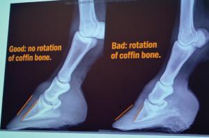
Laminitis is the inflammation of the sensitive laminae. This is what connects the Coffin bone (P3) to the hoof wall. Founder is the mechanical change within the hoof: sinking of the Coffin bone, rotation of the Coffin bone, or both.
Endocrine-related Lamintis
Dr. Melissa Hines followed, describing Endocrine-related laminitis. She explained the Pituitary Pars Intermedia Dysfunction (PPID, Equine Cushing’s Disease) and Equine Metabolic Syndrome, and how both are related to laminitis.
Colitis-related Laminitis
Dr. Megan McCracken covered Colitis-related Laminitis. Colitis is an inflammation of the large bowel that includes the cecum and large colon. There are multiple causes, both infections and non-infectious. The main clinical sign is severe diarrhea, which causes significant problems including excessive fluid loss and dehydration. The heart has more difficulty in pumping the blood, which is thick or sludgy, so less oxygen transported to all parts of the body; lack of oxygen can cause cell death.
Other consequences include secreting large amounts of fluid and protein into the bowel lumen; reduced absorption of water, electrolytes, and nutrients; and the mucosa of the inner most layer of the colon wall becomes “leaky.” As this happens, bacteria and their toxins can move out of the hind gut lumen and into the blood stream, whereby colitis becomes a whole body disease, causing sepsis and endotoxemia. This, then, kicks in the body’s immune system.
Sepsis is the body’s extreme response to bacterial infection. Endotoxemia is the body’s extreme response to a certain part of the cell membrane of Gram negative bacteria. The horse’s body responses to sepsis/endotoxin result in massive inflammation, which kills the bacteria, but also causes significant collateral damage to the body.
Sepsis and endotoxemia are the components of colitis that can lead to laminitis. Current research looks at increased inflammatory mediators in the lamellae and the failure of the epithelial cell adhesion molecule that is responsible for holding the lamellae together. If the lamellae are not holding, then laminitis and effects on the coffin bone rotation occur.
One of the common treatments applied for laminitis is cryotherapy or icing. A landmark study by Van Eps and Pollitt (2004, 2006) showed that horses could tolerate long periods of hypothermia (having their leg immersed in ice water). Ice therapy decreased the blood flow and resulted in decreased inflammatory mediators in the foot and decreased laminitis development. It revolutionized treatment of sepsis and endotoxemia associated laminitis.
More recent research by Van Eps, et al. (2014) further looked at the effects of hydrotherapy (ice water) on sepsis/endotoxemia induced laminitis and found less cellular damage and lamellar separation in the iced limb. However, the disadvantages to using ice boots are that they’re very labor intensive, require 24-hour care, and the horse must tolerate the ice boots. Treatment must also include other therapies that address the sepsis/endotoxemia and related dehydration, poor oxygen circulation to decrease the potential for laminitis.
Treatments include mechanical support, such as Soft Ride boots, IV fluids, antibiotics, and anti-inflammatory medicines. In conclusion, Colitis is a serious, potentially life threatening disease. It leads to sepsis/endotoxemia, which leads to laminitis.
Laminitis from a Surgical Perspective
Dr. Ellie Cypher explained laminitis from a surgical perspective. Her concerns were morbidity and mortality of the horse and long term complications and management.
To reiterate: laminitis is the inflammation and dysfunction of the laminar epithelium. Anatomy consists of the epidermal hoof and all its connective tissues; cartilage, digital cushion; navicular bone, P3 (coffin bone), and distal portion P2; associated ligaments, tendons, and synovial structures. All these are continuous with the epidermis of the coronary band.
The laminae, working like Velcro, hook the Coffin bone to the hoof wall. At the same time, the deep digital flexor tendon and superficial flexor tendon are pulling the bone away from the hoof wall.
Causes of laminitis include sepsis, metabolic/endocrine causes, and mechanical causes: laminitis of the unaffected supporting limb as a result of a painful condition, such as fractures and joint infections (as happened with Barbaro). A good deal of the rest of her presentation covered this support limb laminitis. To illustrate the results of this type of laminitis, she showed radiographs of hooves with laminitis with rotation and sinking. Some of the radiographs showed drastic changes in the horse’s hoof within just one week! Severe rotation can occur, but with sinking, the whole bony structure is pushed down to the sole. In this situation, the prognosis is poor.
Another diagnostic tool is venogram, which measures the blood flow to the foot.
Treatment starts with resolution or control of the primary condition. Treatment continues with medications and diet changes. Systemic treatment includes icing in the early stages and laminar support (Soft Ride boots, deep sand bedding, and wedges/pads).
The one surgical option should be a last resort, a salvage procedure: a tenotomy. The aim is to reduce shearing stresses on the dorsal lamella. The deep digital flexor tendon is surgically cut mid-metacarpal. Post-operative treatment lasts about 6-8 weeks so that, hopefully, a normal hoof-pastern axis is possible. The survival rate of the horse is tentative, with only 59% at 2 years.
Introduction to Tribute Equine Nutrition
Tribute Feed nutritionist Nicole Rambo, Ph.D. explained how quality nutrition leads to healthier horses and better performance with less feed and fewer supplements. As a horse owner herself, she recognizes the importance of and results from good quality balanced horse feed. Tribute Feed is a family-owned company, Kalmbach, based in Ohio since 1963. The company first formulated horse feeds in 2004.
What constitutes quality feed, she says, are: fixed formulations, a balanced animo acid profile, organic trace minerals, antioxidants, pre/probiotics and digestive enzymes, Omega-3 and Omega-6 fatty acids, formulations with added Glucosamine and MSM.
Nutrition begins with forage first: legume and/or grass forage. Then consider a horse’s special needs, like metabolic conditions, obesity, hyperactivity, digestive challenges, muscle conditions, and breeding.
She explained that Tribute is a nutrition company, not just a feed company, with the main operation located in Upper Sandusky, Ohio. Horse feeds are made in a separate feed mill, which is ionophore free. Ionophores are commonly added to feed for cattle to improve weight gain and are used in poultry and goat rations as a coccidiostat to control protozoa infections. Ionophores function by increasing cell membrane permeability. But horses are much more sensitive to ionophore poisoning than other species. Ionophore toxicity inhibits sodium and potassium ion transport across cell membranes, which can kill cells—especially muscle cells—leading eventually to total system failure and death. Signs of ionophore poisoning in horses includes poor appetite, diarrhea, muscle weakness, depression, wobbling, colic, excessive urination, sweating, lying down and sudden death.
Rambo emphasized the fact that Tribute feeds use fixed ration formulas, meaning the ingredients and the amounts do not change; all bags have exactly the same ingredients in the same proportion. Compare feed tags with non fixed formula feeds, she said, which use broader terms in describing their ingredients. The ingredients can change because feeds are mixed on a “least cost” formula.
Robert Step was also on hand, representing Tribute Feeds and Knox County Co-Op, to answer feed and nutrition questions.
After lunch, the focus turned to treatment and management of the laminitic horse – first with barefoot trimming and then with shoeing.
Managing the Laminitic Horse Barefoot
Dr. Neal Valk holds a DVM from the University of Tennessee, a certificate from the International Veterinary Chiropractic Association, and certification in natural hoof care and acupuncture. He says the key to managing laminitis is to first identify the triggers (causes). “The horse has an innate ability to heal and we try to optimize that ability to heal,” he said.
He first was introduced to natural hoof care in 2004 and, in his presentation, showed photos of wild horse feet, that are rounded with toes short, heels low, hoof walls straight, and who live in a rough, stony environment. These wild horses spend a lot of hours per day traveling to find food.
In explaining the difference between founder and laminitis, he used the analogy of hitting one’s fingernail (accidently) with a hammer. This is laminitis – getting a hematoma under the nail. If the nail comes off the nailbed, this is like founder. In the worst case of founder, a horse can slough off a foot.
The results of founder on the coffin bone can be: (1) rotate; (2) sink; or (3) rotate and sink.
Valk explained that Jaime Jackson, father of natural hoof care, said treating laminitis is analogous to a snake shedding its skin. As new hoof growth comes out, it will be in alignment with the coffin bone (however it has rotated). “So we mechanically optimize the hoof through trimming, and then wait for the hoof to heal,” Valk said.
While the goals for the patient are always the same, there are different ways to treat laminitis. He has not found just one good way that always works.
He argues that shoes are peripheral loading devices, i.e., they make the horse bear weight entirely on the hoof wall, which is not a good thing with laminitis. Rather than being forced to suspend all of the body weight by laminar attachment, he wants to spread the load to the rest of the hoof – not just the hoof wall, but also the bars, frog, etc. His approach is to unload the hoof wall; unload the laminar attachment and let it heal.
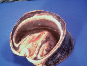
Looking at the bottom of the foot, the white line – juncture of the wall and sole – is a good place to see signs of normalcy or laminitis. If there is a stretched white line, this is a sign of laminitis and a slowly rotating coffin bone. One can also see changes in the hoof wall angle as new hoof grows out above the fever lines.
He doesn’t agree in the surgical option – severing the deep digital flexor tendon. He says this is the one case that allows the coffin bone to move forward.
Valk went on to describe the various types of boots and footing for hoof protection. When a horse wears hoof boots, he wants the horse to spend at least 2-4 hours a day in deep pine shavings. They dry out the moisture that accumulates in boots and the pine oil is therapeutic. With acute laminitis, use ice and/or medication to relieve pain.
Alternatives to boots include casting, clogs, and deep sand footing. Pea gravel is also good because it conforms to the hoof and has a “massage” effect, stimulating hoof growth.
For recovery and rehabilitation, “movement is life,” he says. Get the pain under control, and then get the horse moving. He is not a big proponent of keeping a horse in a stall long term. He believes giving the horse freedom to move at will is a better option.
The last part of his presentation was on maintenance, feed changes. “Remove the triggers, trim the foot, and provide protection” for the hoof. “At the end of the day, it’s all about the quality of life for the horse.”
Managing the Laminitic Horse With Shoeing
Jeremy Davis is a Certified Journeyman Farrier, has achieved all the top qualifications for a farrier, and is the resident farrier at UTCVM. To treat the laminitic horse, understanding the structures and their function in the foot is very important. Treatment includes: frog support, hoof capsule stabilization, removing pressure from the toe region, trimming to minimize stress on the Deep Flexor Tendon, trimming to help realign the Palmar angle and Coffin bone, and protecting the sole of the foot.
He covered the three phases of laminitis: acute, sub-acute, and chronic.
Acute stage is the onset of laminitis, where one sees he horse in the rocked back stance. The sub-acute phase is when the majority of the changes to the coffin bone occur; the horse has a bounding pulse and often an infection in the foot due to bacteria. The tearing apart of the white line and the laminae occurs. In this stage, monitor the digital pulse closely. In the chronic stage, most of the pain has subsided, rotation has taken place, and the foot starts to show changes in appearance and shape. Founder rings start to appear. One can see misshapen hooves and founder rings.
In the acute phase, the shoer is very conservative. The veterinarian is heavily involved to determine the cause(s), get the horse stabilized, and utilize medications and cryotherapy (cold therapy). “The farrier can’t prevent or stop changes within the foot until the horse’s system is stabilized,” he said.
Several options in this stage include: packing with a foam board in the caudal aspect of the foot; lily pads; Soft Ride boots; and a stall with deep sand. “Shavings don’t offer as much support as sand; it’s firm but compressible footing.
In the sub-acute stage, the horse will be lying down a lot, he said. This good, and if the farrier can do the work while the horse is lying down, that’s better.
The chronic phase is where most of the farrier work takes place. He explained and showed examples of the various types of shoes a farrier uses, beginning with the W shoe and the alternative Clog for frog support. Another type of shoe minimizes the stress on the deep flexor tendon. It transfers weight bearing to the superficial flexor tendon and suspensory ligament. He also uses a rail shoe, a rocker/roll shoes, depending on what works for the horse. He has done a lot of the same things with clog shoes. The goal is protecting the thin sole. And before any type of shoe is placed, the trim must be absolutely correct.
To finish the day, Nicole Rambo, Ph.D. with Tribute Feeds was back with a presentation on Feeding for the Ideal Body Weight. Dr. Diana Unzaga gave a Case Presentation. And after all the presentations were completed, attendees could tour the UTCVM facilities. All in all, there was plenty of sound information, lots of food and refreshments, and a delicious lunch.
