By Nancy Brannon, Ph.D.
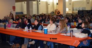
The University of Tennessee College of Veterinary Medicine (UTCVM) offered a wealth of information about horse care at the annual Horse Owners Conference at the Knoxville, Tenn. campus on March 4, 2017. This year’s conference was attended by nearly 90 people and was held in the Equine Performance and Rehabilitation Center, part of the new UTCVM facility on the Agricultural Campus on River Drive. Theme of this year’s conference was Rehabilitate, Recover, Renew.
The conference started at 8 a.m. and concluded with a tour of the Equine Performance & Rehabilitation Center from 4:45 to 5:30 p.m.
The conference is designed for both those who have owned horses for years and those who are new to the horse world. Conference speakers included board-certified faculty of the UT Department of Large Animal Clinical Sciences, UT Extension Equine Specialist, and the veterinary college’s Certified Journeyman Farrier. Topics included general introduction to body systems, lameness, hoof care, neurologic conditions, performance horse nutrition, stretching and warming up exercises, introduction to regenerative medicine, environmental enrichment and strengthening, and proprioception exercise demonstration. Veterinary professionals could gain up to 8 hours of CE credit.
In addition to the lecture presentations, there were plenty of handouts of conference materials for participants to take home.
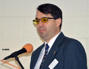
Dr. Steve Adair was the first speaker with a general introduction to body systems and how that may be related to poor performance. “Abnormalities in any body system or location can result in poor performance and any horse can be affected,” he explained. “The most common systems involved in poor performance are the Musculoskeletal, Respiratory, Cardiovascular, Gastrointestinal, and Mental/behavioral.” Adair then gave a more detailed explanation of each system, with specific examples of several types of performance horses and the specific issues involved. This general overview was continued in other presentations on specific systems.
Dr. Jose Castro was next up with a detailed presentation on lameness, including how to conduct a lameness exam, and causes and consequences of a variety of types of injuries. Very helpful were videos of abnormal movements in horses with Dr. Castro’s explanation of how to tell on which leg the horse is lame. For example, in front leg lameness, the horse’s head bobs down on the sound side. In hind leg lameness diagnosis, look for asymmetrical pelvic movements, or the “hip hike;” the lame hind leg goes up.
He went over each stage of the lameness exam and then explained common conformation faults and their resulting effects of way of going. In addition to the physical exam, he explained other diagnostic tools, such as Imaging CT and 3D scans, Xrays and Ultrasound. He explained the differences in the amount of detailed information each machine can give.
Next were descriptions of common causes of lameness, where they are located, and typical symptoms and information revealed by the diagnostic tools.
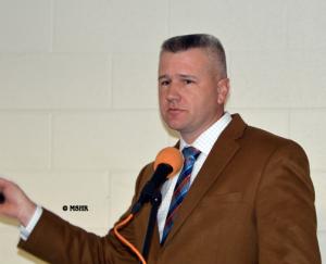
Jeremy Davis, CJF, the resident farrier and head of the Podiatry Program at UTCVM, spoke next about hoof care. Davis has been a farrier for 18 years. “It takes an average of 10,000 feet for a farrier to gain the skills to trim a hoof to a consistent standard,” he told the audience. “It takes 3 to 5 years for a farrier just starting to hone his skills. There are about 50,000 horse shoers in the U.S., and for everyone who goes to farrier school, less than 5% are still shoeing horses at the end of one year and less than 1% are still shoeing after 5 years. There’s a high turnover rate!”
What are the optimal conditions for a farrier to do his best work on your horse? A workplace that is clean, well-lit, dry, level, and, of course, a well-behaved horse.
He discussed whether and when “to shoe or not to shoe.” Begin with the question, what does your horse need? Why shoe? For protection, traction, gait enhancement, and therapeutic purposes. For protection, the examples he gave were trail riding on rocky ground and endurance riding. For traction, his examples included pulling, e.g., a logging horse, and a 3-day eventing horse – shoes with interchangeable studs. For gait enhancement, people automatically think of the padded Tennessee walking horse, but reining horses need sliding plates to get those sliding stops. Therapeutic uses include for founder: a natural balance shoe, a leather pad to protect the sole, and packing to support the frog and internal structures.
Regarding healing hoof injuries, he explained that for the amount of time it took to create the problem in the first place, it takes twice that amount of time to fix the problem. His take away was to evaluate your own horse’s conditions and take care of your own horses according to their needs. “Don’t follow the crowd,” he said. Take into consideration where and how much you ride, talk to your veterinarian and farrier, and then make an informed decision about what your horse needs.
Melissa Hines, DVM, Ph.D., Professor of Equine Internal Medicine and Neonatology at UTCVM spoke about Tying Up. Why do horses tie up? There are two kinds of tying up: sporadic and recurrent. In sporadic tying up, the cause is likely overexertion. In recurrent, there is an underlying disorder.
With recurrent Rhabdomyolysis, which is due to a direct or indirect muscle injury, the causes may be genetic defects, myofibrillar myopathy, or PSSM (polysaccharide storage myopathy), which is a glycogen storage disorder. There are type 1 and type 2 PSSM. Type 1 is caused by a mutation in an identified gene. In type 2, the gene has not yet been identified. Genetic testing is available for type 1 PSSM, through AQHA. AQHA offers a 5-panel genetic test for PSSM, HM, HyPP, GBED, and HERDA (a skin disease). A muscle biopsy is recommended for diagnosis of type 1, and required for diagnosis of type 2 PSSM.
How can horse owners manage this disease? Mainly through diet. Limit carbohydrates and increase fat; balance the vitamins and minerals in the diet. The basis for the diet is high quality grass or oat hay. Avoid grains, sweet feeds, and treats. Add fat through rice bran, canola oil, or flax. Regular, consistent exercise and turnout is the important complement to diet management.
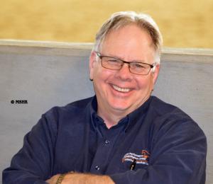
Jim Schumacher, DVM, MS, DACVS, Professor of Equine Surgery at UTCVM, discussed cleft palate and dorsal displacement of the soft palate (DDSP) in horses. An interesting observation is how the horse breathes: when the forefeet leave the ground, the horse inhales; when the forefeet touch the ground, the horse exhales.
Clinical signs of dorsal displacement of the soft palate include: horse suddenly “chokes down (up) during, typically, the latter stages of a race; horse makes abnormal, loud expiratory noise; horse loses coordination of stride; cheeks may bulge; horse may cough, salivate, and swallow frequently.
“Diagnosis includes dynamic endoscopy – on a treadmill or “over-the-ground endoscopy.”
Treatment involves alleviation of any primary disease of the respiratory tract or oral cavity that could cause DDSP, such as small airway inflammation. If the horse is unfit, increase the level of fitness. Correct any dental problems. May need to change the bit, e.g., to a rubber snaffle, or go to a bitless bridle, so the horse can maintain the air-tight seal of the lips. Two types of nose bands may help: an Australian nose band that holds the bit high in the mouth, and a dropped nose band that keeps the horse from opening the mouth. A common technique used in race horses is tying the tongue. The tongue is pulled forward and tied to the lower jaw. This reduces retraction of the tongue and elevation of the soft palate, but does not improve airway mechanics in normal horses.
He discussed other types of treatment, such as sclerotherapy, staphlectomy, sternothyrohoidectomy, thermocautery of the soft palate, epiglottal augmentation, tracheostomy, all of which are of highly questionable efficacy, and the tie-forward procedure, which may be effective in ~80% of horses. This common procedure replaces the function of the thyrohyoideus muscles to prevent DDSP.
Recurrent Laryngeal Neuropathy is also called “roaring,” and is a permanent dysfunction of the laryngeal muscles innervated by the recurrent laryngeal branch of the vagus nerve. It almost invariably involves the left side. This syndrome results in compromised athletic performance through hypoxia. This syndrome has been recognized since the 1800s, but is prevalent in only about 2% of the horse population, mainly in draft horses. Interestingly, women can hear the noise better than men. Also interesting is that horses over 16 hands are most susceptible and is it uncommon in horses <15 ½ hands.
Again, diagnosis is by endoscopy during exercise on a high-speed treadmill or dynamic endoscopy. Schumacher discussed the various surgical procedures used in treatment, with prosthetic laryngoplasty the preferred treatment for recurrent laryngeal neuropathy.
After lunch, Dr. Jennie Ivey, UT Extension Equine Specialist, discussed nutrition for performance horses. The horse has athletic ability that surpasses other animals of similar body size. As an athlete, the horse has many physiologic attributes: high maximal aerobic capacity, large intramuscular stores of glycogen; high mitochondrial content, increased oxygen-carrying capacity of the blood; efficiency of gait; and efficient thermoregulation.
In determining appropriate diet and nutrition for one’s horse, the first considerations are how hard is the horse working and does the diet supply sufficient nutrients for the workload? Light work is done by most trail and arena performance horses; moderate work includes timed events, jumping, and cattle working events; horses with heavy workloads are race horses, cutting horses, and polo ponies.
Next in consideration is aerobic (with air) vs. anaerobic metabolism, each of which utilize different energy substrates. Long, slow distance work utilizes fats. Short, high-velocity/strength work requires simple carbohydrates.
Digestible energy and crude protein requirements increase from maintenance through light, moderate, and heavy exercise.
Feeding fundamentals: (1) feed forage first; 2-2.5% of bodyweight per day; (2) concentrates, appropriate for age and stage of life; no more that 0.75% of body weight per feeding; (3) supplementation only if feeds do not meet nutritional requirements by diet alone.
Ivey discussed the proper way to increase carbohydrates in the diet; which horses may need “low starch” diets; how to add fats to the diet; and feeding proteins, emphasizing quality over quantity. For maintenance, horses need ~8% crude protein in their diet.
For vitamins and minerals, she said usually both are met by increased feed to meet energy needs. The Calcium to Phosphorus ratio should be about 1.5:1 for maintenance, although growing horses in training may need more calcium.
Something to remember about supplements is that the contents are not regulated and many are not tested by research. Ivey said most horses do not need additional supplements if they have well-balanced diets.
However, she discussed supplements for horses with exertional rhabdomyolosis in heavy exercise. These horses may need more Vitamin E (2500-5000 IU) to reduce muscle membrane leakage, and the E should be fed in conjunction with Vitamins C and/or A.
What about electrolytes? Ivey says they are only needed if the horse is heavily sweating.
Joint supplements: not all horses respond to them because many do not make it into the bloodstream in sufficient quantities. Typical ingredients include glucosamine, chondroitin sulfate, MSM, and hylauronic acid.
Hoof supplements include biotin with B vitamins, which activates keratin production; iodine, which aids in thyroid hormone production for tissue development; and zinc for hoof health and metabolic function.
How do you monitor your horse’s fitness? One way is by measuring the recovery heart rate (HR). First, determine the normal heart rate for your horse, about 28-40 beats per minute. Then after work, monitor the time it takes to get back to resting HR. If the HR returns to normal after 15 minutes post-exercise, the horse has worked adequately to maintain its current level of fitness. If HR recovery occurs after 30 minutes, the horse has been adequately stressed to increase fitness. If HR recovery takes longer than 30 minutes, the horse has been worked too hard. It’s time to scale back the intensity or duration of the work.
Another way is to measure the horse’s respiratory rate recovery. Normal resting respiration rate is 8-16 breaths per minute. The heart rate to respiratory rate should be 3:1 to 2:1.
Next on the agenda was Steve Adair, MS, DVM, DACVSMR, Director of Equine Performance Medicine and the Rehabilitation Center at UTCVM, with a demonstration of Equine Baited Stretches, aka Equine Carrot Stretches. The “bait” commonly used is carrots, long strips of carrots so one can keep the fingers out of the way! These stretches are best done when the muscles are warm, i.e., after exercise, and increase flexibility, core strength, and balance. Two to three repetitions of each stretch are recommended (10 second hold for first and second; 20 second hold for last).
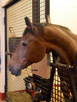
Adair showed how to do various stretches on Camilla, a 28-year-old horse who looks really good for her age! He showed the poll extension, cervical extension, poll flex, mid-cervical flex, lower cervical flex, poll bend, mid-cervical bend, lower cervical bend, butt tuck, butt tuck with side bend, sternal lift, front flexor stretch, front extensor stretch, hind flexor stretch, hind extensor stretch, and hind flexor stretch. His instructions for each included “be careful!”
“All this is passive stretching,” he explained, as opposed to being in the saddle and pulling the head around where the horse gives some resistance. In passive stretching, the horse is doing it on its own and is relaxing in the stretch. “Make sure you do equal stretches on both sides.”
Dr. Adair is a Certified Equine Rehabilitation Practitioner, and he clarified what a chiropractor does. “Chiropractors fix abnormal joint motion. They don’t put bones back in place. They do physical therapy and restore normal joint motion.” Dr. Adair’s presentation included a handout in the packet about doing equine carrot stretches.
Dr. Adair continued with a presentation on Equine Regenerative Medicine, which is a strategy for replacing, repairing, or enhancing the biologic function of damaged tissue by means of autologous or allogenic cells. Adair discussed in depth the four types of regenerative medicine: (1) scaffold therapies; (2) growth factor therapies; (3) cellular therapies; (4) autologous conditioned serum (ACM), and the appropriate applications of each.
The day’s lectures concluded with Dr. Tena Ursini, who is a medical resident in the Large Animal Clinic, discussing “Environmental Enrichment – Keeping Your Horse Happy While Out of Work.”
The rest of the afternoon was spent in demonstrations, with Dr. Ursini showing strengthening and proprioception exercises. Then attendees got a tour of the UTCVM Equine Performance and Rehabilitation Center.
Coming Attractions
More learning opportunities are coming up at UTCVM. On April 1, 2017 is a special 1-day CE seminar with Jean Luc Cornille, the Science of Motion. The Science of Motion is a new approach to therapy which, instead of treating the pathological changes or damage, addresses the kinematics abnormalities causing the pathological changes. Every horse moves differently and, since none move perfectly, especially with a rider on their back, even minor defects in gait can eventually result in lameness. Careful analysis of how a horse moves and an individualized training program can enhance performance and rehabilitate injuries. Find more information at www.scienceofmotion.com. This seminar will be from 8 a.m. to 5 p.m., with a morning lecture followed by six lessons and demonstrations. Find conference information at: https://vetmed.tennessee.edu/ce/Pages/scienceofmotion.aspx.
On April 8, 2017 UTCVM hosts an Open House from 9 a.m. to 5 pm. Everyone is invited and encouraged to bring the family. This year’s theme is “If the shoe fits…” showcasing the various “shoes” or roles that veterinarians wear/ fill. Find more information at: http://tiny.utk.edu/VETMEDopenhouse.
