
By Tommy Brannon
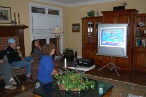
Massar Stables in Arlington Tennessee hosted a special education program for Delta Dressage Association members at the home of Kathy Massey on March 23, 2012. The featured speaker was Dr. Cindy Weis, who practices Veterinary Medicine in Eads, TN and the surrounding area.
Dr. Weis used a slide presentation to educate the members about thermal imaging technology and how it is used to diagnose health problems in horses, as well as saddle fitment and rider position. She said that she has been using her thermal imaging camera for a few years now, and it has turned out to be invaluable.
The images that she displayed on the screen were in the colors that the thermal imaging machine uses to indicate temperatures. The camera registers the skin temperature and can show in increments of five degree Celsius differences. White is the hottest; red is somewhat hot; green, cooler; blue, cold; and black indicates there may be no circulation. Hot spots usually indicate there is a problem either from friction, increased circulation or inflammation. Cold spots can indicate lower circulation or even frostbite. Tendons show up in blue. Red may be normal in many circumstances. The coronet band is red because of the active metabolism to continuously grow a hoof. Laminitis will show up as black in the hoof. Particularly for laminitis, thermal imaging can help one to decide whether the problem is correctable.
A question from the audience asked whether a winter coat may affect the thermal image. Dr. Weis answered that the coat will not, but a drafty barn will. It is important to test where there is no wind.
She said that thermal imaging is a “go to” tool, particularly for diagnosing lameness problems. Its asset is that it is non-evasive.
There are times when the regular rider of a particular horse can tell that “he is just a little off.” Horses will compensate for pain. One part of the body may be in pain, but the horse’s compensation may make one think that the problem is in another part of the body. The lameness may be diagonal. This is one reason why lameness can be very challenging to diagnose. She said it works well in finding stifle issues. “I put the camera on it and it shows right up!”
She discussed several studies that were done at race tracks using thermal imaging. The horses were tested every two weeks, which helped the trainers see problems that could be corrected before they manifested, and before it was too late to correct.
One interesting study with thermal imaging found that some horses use different muscles to swim than other horses. There are about five different styles of horse swimming. This can be important to know when using hydro therapy. One needs to know if it is effective in exercising the proper muscles to get the desired results.
Dr. Weis said that since thermal imaging is a ”go to tool,” it needs to be used in conjunction with other diagnostic techniques such as radio grams (X-Rays), ultra sound, magnetic resonance imaging (MRI), and nuclear scintigraphy (bone scan) to pinpoint the problem. She works with other veterinary clinics, such as Tennessee Equine Hospital and Mississippi State University, that have these diagnostic tools.
Additional topics that were discussed at the meeting were the need for electrolytes, trailering, Cushing’s disease, bran diets, parasite management, trailering and Melanoma in horses.
Delta Dressage Association has informative meeting for its membership as well as well as trainer clinics and shows. The Delta Dressage website is www.deltadressage.com


