

By Nancy Brannon, Ph.D.
Ida Hammer was back at Wildwood Farm in Germantown, Tenn. August 27-29, 2021 for another equine hoof care and trimming clinic. Host Melanie Taylor graciously opens Wildwood Farm to eager students who want to learn all they can about hoof care and how to properly trim horses’ hooves.
“Following the success of the Gold Medal Swedish Show Jumping Team at the Tokyo Olympics, where two of its members competed on barefoot horses, there has been a tremendous amount of interest in going au natural. Ida says that barefoot horses read the ground better because of proprioceptors in the frog and digital cushion,” Melanie Taylor said of the clinic.
The first day was spent covering the structures of the equine hoof, how they function, and the diseases and lamenesses that can result from imbalance, improper nutrition, too early and too intense riding and training, and other factors.
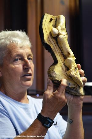
Hammer packs a lot of information into her first day’s presentation, and she shows real examples of equine hoof bones (from cadavers) from her extensive collection so one can physically see what they look like up close, how they fit together, and the variety of changes in bone that can cause problems. She also includes stories of her experiences with her own horses and, with her self-effacing humor, is not afraid to share the mistakes she has made so that all can learn from them. “You do dumb things until you know better,” she quipped. She also does funny imitations of horses’ behavior. Some of the stories of lameness issues are written on her website: https://mackinawdells2.com/about-me.html.
How did she get started on the “barefoot” trimming road? It was her cutting horse and her blue roan Hancock Foundation Quarter Horse named Bobbi that led her to seek out Pete Ramey and Jaime Jackson to help these horses who diagnosed with navicular disease, and a prognosis that one wouldn’t make it to her teenage years. She began learning about Jaime Jackson’s approach to the Natural Trim and Pete Ramey’s Care and Rehabilitation of the Equine Foot. She “joined the AANHCP (Association for the Advancement of Natural Horse Care Practices) and traveled the U.S. learning from the best of the best.” She then applied these methods to the horses with navicular disease: “We took their shoes off, trimmed them properly, and the horses were sound within three months.”
Over the years she has worked directly and indirectly with Pete Ramey; she has several CE hours with Dr. Robert Bowker of Michigan State University, Dr. Debra Taylor of Auburn University, and the late Dr. Kerry Ridgway; she completed Dr. Eleanor Kellon’s NRC Plus and Nutrition as Therapy.
She now has her own program called the Mackinaw Dells 2 Applied Whole Horse Hoof Care Program and is also a mentor for the Progressive Hoof Care Practitioners (PHCP). For her and her students, learning is a lifetime endeavor: “Education never stops; it’s continuous and I’m always learning.” And through her clinics she is “creating a network of knowledgeable people.”
Every horse person knows the adage “no hoof, no horse.” She expands that theme to include “know hoof, know horse.” The most important thing to remember is that “Everything shows up in the feet. Everything that affects the feet affects the body, and everything that affects the body affects the feet.” And it’s of ultimate importance that any hoof trimmer or farrier understand the horse’s anatomy, especially the feet and legs. But even if you don’t plan to be a trimmer or a farrier, knowledge of the horse’s anatomy is crucial to providing better care for our horses.
Hoof as Blood Pump
First and foremost, the horse’s hoof is a blood pump; it is a highly vascular construction and pumps blood like the heart pumps blood. It is filled with veins and arteries, and when the horse loads the hoof, it expands with blood and then when it retracts, a vacuum forms and blood is pumped to the whole body. It’s what Hammer said Dr. Robert Bowker calls the “theory of haemodynamics.” His study of the heomodynamic mechanism of the hoof (updated April 28, 2020) illustrates how the hoof acts as a pump and a hydraulic dampener. [see https://www.theequinedocumentalist.com/post/haemodynamic-mechanism-the-key-to-hoof-health] Editor’s note: Also see Anne Riddell’s article, “Heart of the hoof,” at Equine Wellness: https://equinewellnessmagazine.com/heart-hoof/
Bowker suggests, “When the foot hits the ground, the bars of the heels and pillars of the hoof wall force a small ‘shelf’ of the cartilage outward, creating negative pressure in the digital cushion. Impact is thus transmitted to a complex venous network inside the cartilage, creating more negative energy, which draws blood up from the solear area of the hoof.” (Radiograph from University of Georgia, Dr. Randy Eggleston)
Hammer showed a live-loop video of hoof movement and blood flow in the hoof so folks could see how this pump mechanism works in real time. This was followed by a color photo showing the vascularity of the hoof and how the coffin and navicular bones of the hoof are “bathed” in enriching blood flow.
While she is not opposed to shoeing horses, Hammer talked about the effects of nailed on shoes on the ability of the horse’s hoof to expand and contract properly to pump blood. She also showed a comparison video of a shod horse moving simultaneously with a barefoot horse. The slow motion showed all the vibration and shock from landing that moves up the horse’s leg in the shod hoof, but not so much in the barefoot horse. She said that when a horse is wearing the “performance shoe,” there is even less shock and vibration going up the horse’s leg than on a barefoot horse. She explained how this vibration going up the horse’s leg can break down ligaments.
Coffin bone
“All hoof trimmers must understand the horse’s distal anatomy,” she emphasized. The furthest point in the horse’s distal anatomy is the coffin bone, also called P3 or the pedal bone. She showed various examples of the coffin bone, how P2 fits the coffin bone, what sidebone looks like, a hoof with ringbone, and one with both sidebone and ringbone. She showed the vascular holes that allow blood flow through the bone and the crena, or cleft in the front of the coffin bone. The crena is a shallow notch at the middle front of the coffin bone that can be of different sizes and might or might not extend to the outer hoof wall. Every coffin bone has one, she said, and some are so small as to be almost imperceptible. But some horses have an exaggerated crena; why? Theory is that it is caused by shoes with toe clips. She doesn’t necessarily agree with this causal theory, but did say that in a horse with an exaggerated crena, a toe clip can exacerbate problems.
[See hoof anatomy illustration: https://open.lib.umn.edu/largeanimalsurgery/chapter/foot-anatomy/]
She described the coffin bone as the natural arch of the hoof. Every coffin bone has its own unique amount of concavity, so that’s why it’s important not to carve the sole.
Hammer picked up a whole lower limb to show how all the bones attach and where tendons and ligaments attach. Then she showed a slide of the coffin bone with the sole pulled off so folks could see all the blood vessels.
On laminitis: “The number one cause of laminitis is metabolic,” Hammer said. “Horses are foragers, not grazers. Wild horses eat, then move; eat some more, move some more.” In pastures that are groomed for horses, they can get too much sugar from the grasses.
Navicular bone
The navicular bone “is the shape of a little boat,” she described. It is sometimes called the distal sesamoid bone. She pointed out the little pit holes that can cause degeneration of the deep digital flexor tendon. “Little changes in the bone can mean a lot of changes on the tendon,” she explained.
Navicular disease is not just one issue, but there are types of navicular disease: for examples, bone spurs, degeneration, bursitis, and ligament tears. She has learned not to always promise complete healing of a particular problem. “You can make things more functional and comfortable for the horse. You can’t fix everything, but you can manage the problem.
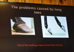
Ida explained how long toes cause problems. “Every centimeter of extra toe length equals 110 pounds of force on acting tendons. Long toes can be a cause of navicular disease. Long toes create hoof distortion and can create problems throughout the body. And we get long toes because every farrier is taught not to cut inside the white line. However, the white line is in the insensitive laminae, so we can cut into the white line to shorten the toe. There is a difference between shortening the hoof and lowering the hoof. By not cutting inside the white line, we often get low feet and long toes, rather than shortening the toe.”
She next turned her attention to X-rays, which are extremely helpful to trimmers and farriers to know what’s going on with the bones. “What do we want to see in X-rays?” First, mark where the coronary band is so we can see the true alignment of the hoof wall and the coffin bone. Both hooves should be at the same block height so we can get true readings of the joint spaces. She also wants the veterinarian to mark the apex of the frog. Get lateral views. A “navicular shot” or “skyline” shows the condition of the navicular bone. To see if the horse has pedal osteoarthritis, get a “solar margin” shot.
Horses all have a wear pattern, and making daily observances of the wear pattern can give indications of any small problems developing before they become major ones.
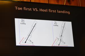
Toe first vs. heel first landings
“An impressive section of her PowerPoint displayed the difference in a toe first versus heel first landing in the normal foot fall of the horse,” wrote Melanie Taylor. The horse should normally land heel first; that keeps all the bones in proper alignment. If the horse lands toe first, that puts the bones in misalignment, which also puts the soft tissue in misalignment. To evaluate how the horse is landing, look at the leg, not the hoof: if the leg is aligned correctly, the horse is landing correctly. But a horse that lands toe first can develop navicular bone problems, since the navicular bone is pushed back and down in a toe-first landing.
Digital cushion
“If the digital cushion is not robust, the whole foot is weak,” Hammer explained. The digital cushion is a wedged-shaped structure with a fibro-fatty composition in young horses, which hardens into a fibrocartilagineous tissue in the adult horse. It is very elastic and has very few blood vessels and nerves.
It is located in a wedged-in position between the lateral cartilages on the side, the deep flexor tendon on the top, and the frog on the bottom and rear. It separates the frog and the bulb from underlying tendons, joints, and bones, providing cushioning protection.
When it is compressed by the pastern bones and frog, it absorbs shock and cushions the bones. As weight is placed on the hoof, pressure is transmitted through the phalanges to the wall and onto the digital cushion and frog. The frog, a highly elastic wedge-shaped mass, normally makes contact with the ground first. The frog presses up on the digital cushion, which flattens and is forced outward against the lateral cartilages. The frog also is flattened and tends to push the bars of the hoof wall apart. When the foot is lifted, the frog and other flexible structures of the foot, such as the digital cushion, return to their original position.
[Source: “Digital Cushion.” 2020. Horses. Jan. 22. https://horses.extension.org/digital-cushion/]
“We can palpate the digital cushion by press and release of the frog,” Hammer explained. “In a young horse that has not fully developed the digital cushion, if we put shoes on them, we suspend the development of the digital cushion. As 2-year-olds, the growth plates are not closed yet – at least not until three years of age. Once we remove the shoes and let the horse use the frog again, the horse can regenerate the cushion, but it’s a slower process.” Hammer referred to Dr. Taylor’s (Auburn University) studies of the development of the digital cushion. Find more information about these studies at: https://www.hoofrehab.com/AuburnUvetschool.htm
“How do you determine if the horse has a good digital cushion? The heel buttresses should be lined up with the middle of the canon bone. The digital cushion has to be in a good place to absorb shock and support the bones. If the heel bulges out behind, the digital cushion is in the wrong place – displaced to the back – and cannot fully support the bones of the foot and leg.” To illustrate, she showed video footage of how the digital cushion supports the coffin and navicular bones as the horse moves and flexes. The video, Extract II of equine Foot Studies by Dr. C.C. Pollitt, can be viewed at:
https://fo-fo.facebook.com/Gwennael.Cadet.Public/videos/extract-ii-of-equine-foot-studies-by-dr-cc-pollitt/1900565936839197/. You will gain new insight into what happens inside the hoof when your horse moves.
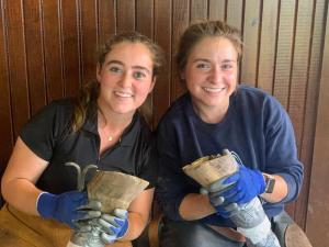
“The second and third days of Ida’s clinic were Beginner and Intermediate Trim. Each participant was given a cadaver leg with hoof to trim so they could practice on the real thing with no concern of doing damage to a live horse. This class is worth taking whether you intend to trim your own horses or not. It gives you better perspective on how the foot should be trimmed through Ida’s demonstrations and dissections, and you begin to develop ‘x-ray vision’ to the inside of the foot,” Melanie Taylor explained.
Read Melanie Taylor’s blog about the clinic at: http://melaniesmithtaylor.com/wildwoods-continuing-education-in-a-nutshell/
Find more information about Ida Hammer’s Whole Horse Trimming at: https://mackinawdells2.com. If you missed this clinic, Exploring the Equine Hoof is now available online at: https://mackinawdells2learning.thinkific.com/
Ida Hammer was back at Wildwood Farm in Germantown, Tenn. August 27-29, 2021 for another equine hoof care and trimming clinic. Host Melanie Taylor graciously opens Wildwood Farm to eager students who want to learn all they can about hoof care and how to properly trim horses’ hooves.
“Following the success of the Gold Medal Swedish Show Jumping Team at the Tokyo Olympics, where two of its members competed on barefoot horses, there has been a tremendous amount of interest in going au natural. Ida says that barefoot horses read the ground better because of proprioceptors in the frog and digital cushion,” Melanie Taylor said of the clinic.
The first day was spent covering the structures of the equine hoof, how they function, and the diseases and lamenesses that can result from imbalance, improper nutrition, too early and too intense riding and training, and other factors.
Hammer packs a lot of information into her first day’s presentation, and she shows real examples of equine hoof bones (from cadavers) from her extensive collection so one can physically see what they look like up close, how they fit together, and the variety of changes in bone that can cause problems. She also includes stories of her experiences with her own horses and, with her self-effacing humor, is not afraid to share the mistakes she has made so that all can learn from them. “You do dumb things until you know better,” she quipped. She also does funny imitations of horses’ behavior. Some of the stories of lameness issues are written on her website: https://mackinawdells2.com/about-me.html.
How did she get started on the “barefoot” trimming road? It was her cutting horse and her blue roan Hancock Foundation Quarter Horse named Bobbi that led her to seek out Pete Ramey and Jaime Jackson to help these horses who diagnosed with navicular disease, and a prognosis that one wouldn’t make it to her teenage years. She began learning about Jaime Jackson’s approach to the Natural Trim and Pete Ramey’s Care and Rehabilitation of the Equine Foot. She “joined the AANHCP (Association for the Advancement of Natural Horse Care Practices) and traveled the U.S. learning from the best of the best.” She then applied these methods to the horses with navicular disease: “We took their shoes off, trimmed them properly, and the horses were sound within three months.”
Over the years she has worked directly and indirectly with Pete Ramey; she has several CE hours with Dr. Robert Bowker of Michigan State University, Dr. Debra Taylor of Auburn University, and the late Dr. Kerry Ridgway; she completed Dr. Eleanor Kellon’s NRC Plus and Nutrition as Therapy.
She now has her own program called the Mackinaw Dells 2 Applied Whole Horse Hoof Care Program and is also a mentor for the Progressive Hoof Care Practitioners (PHCP). For her and her students, learning is a lifetime endeavor: “Education never stops; it’s continuous and I’m always learning.” And through her clinics she is “creating a network of knowledgeable people.”
Every horse person knows the adage “no hoof, no horse.” She expands that theme to include “know hoof, know horse.” The most important thing to remember is that “Everything shows up in the feet. Everything that affects the feet affects the body, and everything that affects the body affects the feet.” And it’s of ultimate importance that any hoof trimmer or farrier understand the horse’s anatomy, especially the feet and legs. But even if you don’t plan to be a trimmer or a farrier, knowledge of the horse’s anatomy is crucial to providing better care for our horses.
Hoof as Blood Pump
First and foremost, the horse’s hoof is a blood pump; it is a highly vascular construction and pumps blood like the heart pumps blood. It is filled with veins and arteries, and when the horse loads the hoof, it expands with blood and then when it retracts, a vacuum forms and blood is pumped to the whole body. It’s what Hammer said Dr. Robert Bowker calls the “theory of haemodynamics.” His study of the heomodynamic mechanism of the hoof (updated April 28, 2020) illustrates how the hoof acts as a pump and a hydraulic dampener. [see https://www.theequinedocumentalist.com/post/haemodynamic-mechanism-the-key-to-hoof-health] Editor’s note: Also see Anne Riddell’s article, “Heart of the hoof,” at Equine Wellness: https://equinewellnessmagazine.com/heart-hoof/
Bowker suggests, “When the foot hits the ground, the bars of the heels and pillars of the hoof wall force a small ‘shelf’ of the cartilage outward, creating negative pressure in the digital cushion. Impact is thus transmitted to a complex venous network inside the cartilage, creating more negative energy, which draws blood up from the solear area of the hoof.” (Radiograph from University of Georgia, Dr. Randy Eggleston)
Hammer showed a live-loop video of hoof movement and blood flow in the hoof so folks could see how this pump mechanism works in real time. This was followed by a color photo showing the vascularity of the hoof and how the coffin and navicular bones of the hoof are “bathed” in enriching blood flow.
While she is not opposed to shoeing horses, Hammer talked about the effects of nailed on shoes on the ability of the horse’s hoof to expand and contract properly to pump blood. She also showed a comparison video of a shod horse moving simultaneously with a barefoot horse. The slow motion showed all the vibration and shock from landing that moves up the horse’s leg in the shod hoof, but not so much in the barefoot horse. She said that when a horse is wearing the “performance shoe,” there is even less shock and vibration going up the horse’s leg than on a barefoot horse. She explained how this vibration going up the horse’s leg can break down ligaments.
Coffin bone
“All hoof trimmers must understand the horse’s distal anatomy,” she emphasized. The furthest point in the horse’s distal anatomy is the coffin bone, also called P3 or the pedal bone. She showed various examples of the coffin bone, how P2 fits the coffin bone, what sidebone looks like, a hoof with ringbone, and one with both sidebone and ringbone. She showed the vascular holes that allow blood flow through the bone and the crena, or cleft in the front of the coffin bone. The crena is a shallow notch at the middle front of the coffin bone that can be of different sizes and might or might not extend to the outer hoof wall. Every coffin bone has one, she said, and some are so small as to be almost imperceptible. But some horses have an exaggerated crena; why? Theory is that it is caused by shoes with toe clips. She doesn’t necessarily agree with this causal theory, but did say that in a horse with an exaggerated crena, a toe clip can exacerbate problems.
[See hoof anatomy illustration: https://open.lib.umn.edu/largeanimalsurgery/chapter/foot-anatomy/]
She described the coffin bone as the natural arch of the hoof. Every coffin bone has its own unique amount of concavity, so that’s why it’s important not to carve the sole.
Hammer picked up a whole lower limb to show how all the bones attach and where tendons and ligaments attach. Then she showed a slide of the coffin bone with the sole pulled off so folks could see all the blood vessels.
On laminitis: “The number one cause of laminitis is metabolic,” Hammer said. “Horses are foragers, not grazers. Wild horses eat, then move; eat some more, move some more.” In pastures that are groomed for horses, they can get too much sugar from the grasses.
Navicular bone
The navicular bone “is the shape of a little boat,” she described. It is sometimes called the distal sesamoid bone. She pointed out the little pit holes that can cause degeneration of the deep digital flexor tendon. “Little changes in the bone can mean a lot of changes on the tendon,” she explained.
Navicular disease is not just one issue, but there are types of navicular disease: for examples, bone spurs, degeneration, bursitis, and ligament tears. She has learned not to always promise complete healing of a particular problem. “You can make things more functional and comfortable for the horse. You can’t fix everything, but you can manage the problem.
Ida explained how long toes cause problems. “Every centimeter of extra toe length equals 110 pounds of force on acting tendons. Long toes can be a cause of navicular disease. Long toes create hoof distortion and can create problems throughout the body. And we get long toes because every farrier is taught not to cut inside the white line. However, the white line is in the insensitive laminae, so we can cut into the white line to shorten the toe. There is a difference between shortening the hoof and lowering the hoof. By not cutting inside the white line, we often get low feet and long toes, rather than shortening the toe.”
She next turned her attention to X-rays, which are extremely helpful to trimmers and farriers to know what’s going on with the bones. “What do we want to see in X-rays?” First, mark where the coronary band is so we can see the true alignment of the hoof wall and the coffin bone. Both hooves should be at the same block height so we can get true readings of the joint spaces. She also wants the veterinarian to mark the apex of the frog. Get lateral views. A “navicular shot” or “skyline” shows the condition of the navicular bone. To see if the horse has pedal osteoarthritis, get a “solar margin” shot.
Horses all have a wear pattern, and making daily observances of the wear pattern can give indications of any small problems developing before they become major ones.
Toe first vs. heel first landings
“An impressive section of her PowerPoint displayed the difference in a toe first versus heel first landing in the normal foot fall of the horse,” wrote Melanie Taylor. The horse should normally land heel first; that keeps all the bones in proper alignment. If the horse lands toe first, that puts the bones in misalignment, which also puts the soft tissue in misalignment. To evaluate how the horse is landing, look at the leg, not the hoof: if the leg is aligned correctly, the horse is landing correctly. But a horse that lands toe first can develop navicular bone problems, since the navicular bone is pushed back and down in a toe-first landing.
Digital cushion
“If the digital cushion is not robust, the whole foot is weak,” Hammer explained. The digital cushion is a wedged-shaped structure with a fibro-fatty composition in young horses, which hardens into a fibrocartilagineous tissue in the adult horse. It is very elastic and has very few blood vessels and nerves.
It is located in a wedged-in position between the lateral cartilages on the side, the deep flexor tendon on the top, and the frog on the bottom and rear. It separates the frog and the bulb from underlying tendons, joints, and bones, providing cushioning protection.
When it is compressed by the pastern bones and frog, it absorbs shock and cushions the bones. As weight is placed on the hoof, pressure is transmitted through the phalanges to the wall and onto the digital cushion and frog. The frog, a highly elastic wedge-shaped mass, normally makes contact with the ground first. The frog presses up on the digital cushion, which flattens and is forced outward against the lateral cartilages. The frog also is flattened and tends to push the bars of the hoof wall apart. When the foot is lifted, the frog and other flexible structures of the foot, such as the digital cushion, return to their original position.
[Source: “Digital Cushion.” 2020. Horses. Jan. 22. https://horses.extension.org/digital-cushion/]
“We can palpate the digital cushion by press and release of the frog,” Hammer explained. “In a young horse that has not fully developed the digital cushion, if we put shoes on them, we suspend the development of the digital cushion. As 2-year-olds, the growth plates are not closed yet – at least not until three years of age. Once we remove the shoes and let the horse use the frog again, the horse can regenerate the cushion, but it’s a slower process.” Hammer referred to Dr. Taylor’s (Auburn University) studies of the development of the digital cushion. Find more information about these studies at: https://www.hoofrehab.com/AuburnUvetschool.htm
“How do you determine if the horse has a good digital cushion? The heel buttresses should be lined up with the middle of the canon bone. The digital cushion has to be in a good place to absorb shock and support the bones. If the heel bulges out behind, the digital cushion is in the wrong place – displaced to the back – and cannot fully support the bones of the foot and leg.” To illustrate, she showed video footage of how the digital cushion supports the coffin and navicular bones as the horse moves and flexes. The video, Extract II of equine Foot Studies by Dr. C.C. Pollitt, can be viewed at:
https://fo-fo.facebook.com/Gwennael.Cadet.Public/videos/extract-ii-of-equine-foot-studies-by-dr-cc-pollitt/1900565936839197/. You will gain new insight into what happens inside the hoof when your horse moves.
“The second and third days of Ida’s clinic were Beginner and Intermediate Trim. Each participant was given a cadaver leg with hoof to trim so they could practice on the real thing with no concern of doing damage to a live horse. This class is worth taking whether you intend to trim your own horses or not. It gives you better perspective on how the foot should be trimmed through Ida’s demonstrations and dissections, and you begin to develop ‘x-ray vision’ to the inside of the foot,” Melanie Taylor explained.
Read Melanie Taylor’s blog about the clinic at: http://melaniesmithtaylor.com/wildwoods-continuing-education-in-a-nutshell/
Find more information about Ida Hammer’s Whole Horse Trimming at: https://mackinawdells2.com. If you missed this clinic, Exploring the Equine Hoof is now available online at: https://mackinawdells2learning.thinkific.com/


