By Lisa Manning, Ph.D. and Marcus Manning
Corneal ulcers can become infected with fungal organisms; called keratomycosisi, this is a common vision-threatening disease of the horse. “Kerato,” means corneal disease, and “mycosis” refers to fungal infection. With all the moisture, heat, and humidity in the mid-south, a fungus can get into a horse’s eye and cause serious infection.
The horse in this case, Cool Beans, aka “Scooter,” is an example of how things can quickly go very badly. Scooter first developed a noticeable pinpoint ulcer, which developed into a larger ulcer, and then a fungus caused it to develop rapidly into an infected corneal ulcer.
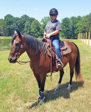
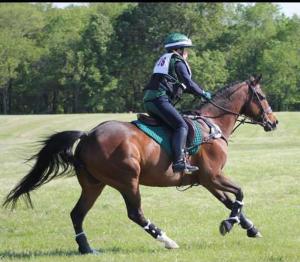
Scooter, a 15-year-old, bay Quarter Horse gelding, came to Missy Carlisle of Coldwater, Mississippi six years ago. Scooter was her eventing horse, who shone in many disciplines including dressage, costume, and ranch riding. Carlisle describes herself “an amateur lady doing this for fun” and her horse Scooter as “unflappable.”
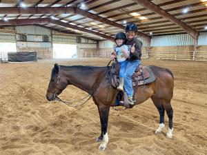
Scooter and Carlisle began Ranch Riding this year at Campbell’s Performance Horse Stables in Moscow, Tennessee with Shane and Catie Campbell.
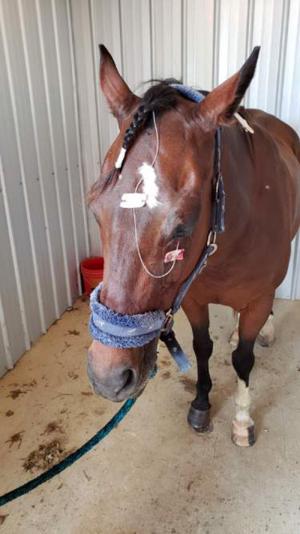
In late May, Scooter was squinting and noticeably uncomfortable from what seemed to be a “scratch” in his left eye. Carlisle wondered if a fly mask may have prevented the injury. She recalled, “When I first noticed Scooter’s eye, I called the vet immediately.” Carlisle’s regular veterinarian, Dr. Megan Hunt, was out of town, so she called Dr. Rilla Reese, of Tennessee Equine Hospital, who came out and started Scooter on antibiotics.
Three days later, there was no improvement, and what appeared to be a scratch grew into a large ulcer. Dr. Hunt placed a subpalpebral lavage catheter to help facilitate easier and more appropriate treatment of the eye. Scooter was started on antifungals, antibiotics, and medication for pain and inflammation. Carlisle and Dr. Hunt agreed that if there was no improvement the following week, they would consult with ophthalmologist Dr. Jane Ashley Huey.
Dr. Huey performed an amnion graft and conjunctival graft, discovering an ulcer that covered 80 percent of Scooter’s eye. Dr. Huey said it was one of the worst cases she had seen. This aggressive eye condition had rapidly worsened from a scratch - to a pinpoint ulcer - to a large, severely infected corneal ulcer in a short period of time.
It was obvious Scooter was in a great deal of pain, despite their efforts to minimize his discomfort. After six weeks of an intense regimen of banamine, antibiotics, and other pain medications, there was still no significant improvement. The symptoms were inconsistent. One day, Scooter seemed more comfortable, then the next day he was clearly in pain.
Dr. Hunt regularly checked Scooter twice a week and tested him for Pituitary Pars Intermedia Dysfunction (Cushing’s disease) to determine if there was an underlying reason that he was not healing. But he tested negative for Cushing’s disease.
At week four, Dr. Huey performed a surgical procedure, removing a brown necrotic plaque from the center of the corneal ulcer. This was a piece of dead cornea that was preventing good drug penetration into the cornea and delaying healing. Antifungal injections were performed throughout this treatment period.
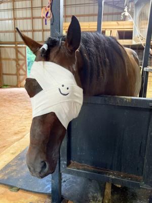
Dr. Hunt continued to check Scooter weekly, who was still showing signs of significant pain, tearing, squinting, and often refusing to open his eyelid. Drs. Huey and Hunt evaluated Scooter together and agreed Scooter was not improving as expected, and both determined the eye should be removed.
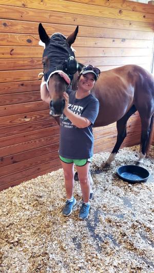
Carlisle explained how she cared for Scooter over the seven-week period, “Scooter lives at home with us and his eye issue happened during the summer. Being a teacher, I have the summers off, so I was able to dedicate the necessary time to his treatment. Scooter was on a strict no sunlight turnout and medicines had to be administered six times a day. Every morning when I called him, he came running to me nickering. Amigo (his donkey pasture buddy) enjoyed this time because he knew treats were involved.”
“We all struggled,” said Carlisle and described Scooter as “a stoic horse, whose personality never changed, and he never got mad”
Ultimately the decision was made to schedule an enucleation procedure to remove Scooter’s left eye. Dr. Hunt assured Carlisle that many one-sighted horses do remarkably well adjusting to blindness and are able to maintain normal jobs successfully.
Dr. Huey, Dr. Hunt, and Carlisle decided to deliberate their decision over the weekend, taking Scooter completely off medications and treatment over the weekend.
On Monday morning, when Scooter was scheduled for the enucleation procedure, he seemed better. Carlisle prayed as she walked into the barn, “God, let him tell me what to do. Please God, give me a sign.” As Carlisle slowly removed his protective mask, Scooter’s eye was not tearing, or watering.
After learning that Scooter was not tearing and had opened his eye, there was some brief doubt if enucleation was the right thing to do. But the decision was made to go ahead with the eye removal. After the procedure, Dr. Hunt dissected the eye and found a large “purulent” (puss-like substance) abscess on the inside of the cornea, involving the inner layer of the cornea, called the endothelium. At that point, Dr. Hunt concluded, “It confirmed it was necessary and could not be resolved.”
After losing sight in his left eye, Scooter’s visual field has changed, and other senses have become heightened. It is important to approach Scooter from the front and speak to him as you approach.
Looking back on lessons learned, Carlisle reflects, “Horse ownership means making a commitment and a lifestyle change. When you have horses, it’s all about loving them through the good and the bad. You experience joy and sorrow, but, for me, the joy outweighs the sorrow. I have a bond with Scooter that involves patience, compassion, kindness, and most of all, trust. In the coming days, weeks, and months, trust is going to be the key to our partnership as we both adjust through this transition period. But I know Scooter will be back to doing what he does best - winning the hearts of everyone he meets.”
Dr. Hunt makes it clear, “What is most important is the quality of life throughout every decision we make.” She praises Carlisle for her quick action to recognize that her horse’s condition was not improving. With the help of insurance, Carlisle could afford every needed treatment in a timely manner.
This is a success story about teamwork and communication among an owner, her veterinarian, and a specialist. Although the outcome wasn’t what they hoped for, Scooter is adjusting well and has a good quality of life. In fact, Scooter already had been adjusting to being partially blind in his left eye over the two-month period. Dr. Hunt remarked, the key to Scooter’s adjustment to being visually impaired “is trulyhis demeanor; he is a quiet, gentle soul.” Carlisle remains hopeful for their future success.
Dr. Hunt explained when there is an abrasion on the cornea, it can become infected with a fungus and can cause a stromal abscess to form. Treatment includes topical antifungal, antibiotics, and anti-inflammatories. In Scooter’s case, all this wasn’t enough to remedy his rapidly deteriorating condition.
When asked, “What are things to look for in preventive eye care for horses? Dr. Hunt replied, “Look for signs of discomfort, such as, discharge, swelling, change in coloration, tearing, or lashes pointed downwards.” Dr. Hunt reflected, “Unfortunately, in Scooter’s case we didn’t know exactly why his condition worsened and went badly quickly, even in the face of very appropriate treatment.”
In terms of prevention, knowing the signs, noticing them, and acting quickly is the best advice for any horse owner. If you and your veterinarian feel that a disease/injury requires an expert opinion, do not hesitate to bring the expert to the case immediately, without delay.
Intended to be a pasture buddy for Scooter, Carlisle adopted “Amigo,” a 10-year-old donkey from Rocking R Ranch and Rescue in Prentiss, MS. Until recently, Amigo and Scooter were indifferent to each other. Now that Scooter lost sight in his left eye, Amigo helps him navigate the pasture and stays close by to look after Scooter. The two are now best buddies.
As Carlisle opens a new chapter, her outlook is, “I know that everything happens for a reason, and I try to find the positive in everything. So maybe during all this I have found my niche of rehabbing sick and injured horses.”
Find more information on equine ocular conditions at these websites:
Brooks, Dennis E., DVM, PhD. Dipl. ACVO. 2002. “Fungal Ulcers in the Equine Eye.” The Horse. June 1. https://thehorse.com/130475/fungal-ulcers-in-the-equine-eye/
Brooks, Dennis E., DVM, PhD, Dipl. ACVO. 2002. “The Dreaded Corneal Stromal Abscess.” The Horse. August 1. https://thehorse.com/130568/the-dreaded-corneal-stromal-abscess/
Brooks, D.E. 2009. “Equine keratomycosis: An international problem.” Equine Veterinary Education. (21, 5) 243-246. https://beva.onlinelibrary.wiley.com/doi/pdfdirect/10.2746/095777309X409929
Galera, Paula D. and Dennis E. Brooks. 2012. “Optimal management of equine keratomycosis.” Dovepress. March 12. https://www.ncbi.nlm.nih.gov/pmc/articles/PMC6065588/
Toler, Erica, DVM and Diane V. H. Hendrix, DVM, DACVO. 2005 “Equine Infectious Keratitis: Diagnosis and Treatment.” Infectious Disease Compendium. (Vol. 27 No. 5, May) https://www.vetfolio.com/learn/article/equine-infectious-keratitis-diagnosis-and-treatment
