By Liberty M. Getman, DVM, Diplomate, ACVS, Tennessee Equine Hospital;
compiled by Nancy Brannon, Ph.D.
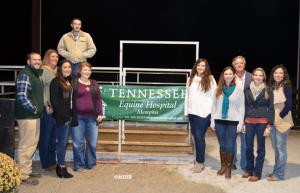
Tennessee Equine Hospital (Eads, Tennessee) held its annual Client Appreciation Party and Seminar on Thursday, November 9, 2017. Once again, guests were treated to a delicious barbeque dinner from Plumpy’s in Arlington, Tennessee, featuring their superb banana pudding! Monty McInturff, owner and main veterinarian at Tennessee Equine Hospital praised the quality of their staff veterinarians, touting them as some of the best graduates from the region’s veterinary schools.
The main speaker for the evening was Dr. Liberty Getman, who explained extensively the process of diagnosing, and the latest advancements in treating, lameness in the horse. Following is a synopsis of her remarks.
When a veterinarian is called to examine and treat a lame horse, Getman explained that the goal is to narrow the source of the problem to a specific area or body part. This process begins with a physical examination, a lameness examination, and possibly a nerve block or a joint block to further isolate the source.
Diagnosis
Diagnostic tools include radiography (X-rays), ultrasonography (or ultrasound), nuclear scintigraphy (bone scan), and magnetic resonance imaging. The nature of the lameness can determine which tools will work best. Getman explained in detail how each works, and what kind of information each captures best.
Radiography is best when looking for bone abnormalities. The black/white images are fairly sharp and can show great detail of bones. But they do not show soft tissues, like muscle, tendons, and ligaments. They also do not work well for larger body parts, such as upper limbs. And the horse would need to be under general anesthesia to X-ray the femur and pelvis. The best uses for radiography are fractures, luxations, bone/joint infections, OCD lesions, arthritis, and laminitis. In her PowerPoint presentation, Getman showed example X-rays of each.
Ultrasonography, aka Ultrasound, works by sending sound waves through the tissue by an ultrasound probe; the probe senses the waves that are bounced back and produces an image. These images are not as detailed as those from X-rays and one has to be “attuned” to interpreting the images. Ultrasound works well for soft tissue injuries like bowed tendons, torn ligaments, and muscle tears; on soft tissue masses like tumors and abscesses; and colic, showing distended intestines, increase in abdominal fluid, and other problems associated with colic.
Nuclear Scintigraphy is also called bone scan or nuclear scan. A radioactive drug is given IV, and it goes to areas of increased blood flow. It can locate arthritis, fractures, infection, and tumors. Radioactivity is detected by a gamma camera, which changes it into a black and white image. The images show a black and white “skeleton,” but are not as crisp/clear as X-rays. But they can indicate areas of increased radiopharmaceutical uptake, aka “hot spots.” Darker areas on the images indicate areas of increased blood flow. Nuclear scintigraphy can image areas of the body that other modalities can’t. It is excellent for bone problems and is more sensitive than X-rays and can image areas that are hard to X-ray such as upper limbs, back, spine, and pelvis. It can show stress fractures. However, the horse must be sedated and remain in the hospital until no longer radioactive.
Magnetic Resonance Imaging, aka MRI, provides multiple cross sectional images of a selected area. The leg is placed in a high powered magnet, which interacts with the H+ (hydrogen) atoms in tissues. It is important that the horse be placed on a special table under anesthesia for this procedure, as the leg must remain perfectly still. The MRI gives excellent definition of soft tissue, better than ultrasound. With so many cross sectional images, the veterinarian can see things that X-rays may not show. The best uses for MRI are for soft tissue injuries of the lower limbs and feet.
Treatment Options
Once the diagnosis is made, there are three categories treatment options available: Medical, Surgical, and Regenerative Therapies.
Medical treatments include shoeing changes, supplements, joint injections, pain relieving medications like bute, banamine, and Previcox®.
Surgical Therapies can be orthopedic or soft tissue surgeries. Orthopedic surgeries include arthroscopy, fracture repair, splint bone removal, and hock arthrodesis (fusion). Soft tissue surgeries are used to treat suspensory injuries, club feet, SDFT injuries, locking patellae, and navicular disease/foot pain.
Arthroscopy, a camera and instruments placed into the joint through small incisions, is used to remove bone fragments, chips, OCD fragments, remove damaged cartilage and soft tissue.
Regenerative Medicine is the most advanced treatment procedure and there are four types: II-1 Receptor Antagonist Protein (IRAP); Platelet Rich Plasma (PRP), Bone Marrow Concentrate (BMAC), and Mesenchymal Stem Cells (MSCs).
IRAP stands for Interleukin-1 Receptor Antagonist Protein. The process uses the horses blood, incubated with glass beads and centrifuged. Once processed, it is injected in the joint where it increases the amount of anti-inflammatory proteins. IRAP can be used to prevent arthritis, after trauma and after surgery.
PRP is Platelet Rich Plasma. This utilizes the patient’s blood plus an anticoagulant. It is centrifuged to concentrate the numbers of platelets and growth factors. PRP is used to stimulate healing in tendon and ligament injuries, cartilage, wounds, and stifle (meniscal injuries). It speeds healing and heals with better tissue, thus reducing scar tissue.
ProStride is a product that combines IRAP and PRP. It uses the horse’s blood with is centrifuged and then injected into the joint. This is done as an outpatient procedure. ProStride decreases inflammation and stimulates healing in tendons, ligaments, and meniscus. ProStride is used for joint injections to treat arthritis, for “maintenance” injections to treat soft tissue damage in and around joints. The TEH veterinarians believe it is safer and healthier for the joint than repeated steroid injections. It does no cartilage damage and can be used in young horses. Its effects can last approximately a year.
BMAC is bone marrow concentrate. This procedure utilizes bone marrow taken from the sternum or hip, which is then centrifuged. The process increases the number of stem cells and growth factors. Bone marrow injections can promote normal healing of tendons and ligaments in the treatment of tendonitis and suspensory injuries.
MSCs, i.e., Mesenchymal Stem Cells are harvested from bone marrow, sent to the lab to be grown in a culture, and then sent back to the veterinarian in large numbers of MSCs. A veterinarian can even use stem cells banked from another horse. The beauty of stem cells is their transformative ability to become a wide variety of different kinds of tissue types.
MSCs are used to promote normal healing of tendons and ligaments – they heal quicker and with better quality tissue, i.e., reduced scar tissue. MSCs can be used to treat laminitis. They are injected directly into the damage structure or regional limb perfusion – a technique with provides a high concentration to the soft tissues, joints, and bones of the limbs.
For more information about any of these treatment procedures, contact your veterinarian and/or Dr. Liberty Getman at Tennessee Equine Hospital in Thompsons Station, TN: (615) 591-1232 or email: lgetman@tnequinehospital.com.
