
Mark A. Akin, DVM
Today’s equine athlete is asked to perform at higher and higher levels. Despite great strides in Veterinary Medicine, nutrition, farriering, and conditioning, today’s equine athlete can become victim to subtle injury which can cause lameness, poor performance, or both. This article takes on three of those often overlooked causes of lameness and poor performance in an attempt to put them in perspective.
A Sacroiliac desmitis and arthropathy
B Trochanteric Bursitis
C Cervical Arthropathy
Sacroiliac Desmitis and Arthropathy
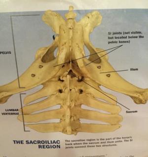
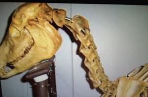
Anatomically the SI region pain is where the ilium (part of the pelvis) and the sacrum (part of the vertebral column) connect:
The Sacroiliac (SI) is often the culprit as it pertains to lower back issues, lameness and poor performance in both the English and Western performance horse. These SI can be primary or can be commonly secondary to lower limb issues concerning the hocks and stifles.
Clinical signs of sacroiliac disease in the performance horse include but aren’t limited to:
-poor performance
-unwillingness to go forward
-a refusal to “collect”
-pain on palpation of the lower back and croup (sacral tuberosity)
-kicking out when trotting or cantering
-poor attitude during exercise, just “crabby”
-frequent shifting of weight
-lack of desire to have rear flexions performed
Treatment involves multiple modalities and should be done in concert with each other. My typical treatment plan involves the following and can last from three weeks to 3 months.
-Rest. The cornerstone of any rehabilitation program.
-Controlled exercise program which involves a lot of lunging which leads to work under saddle. Starting with easy tasks for the horse and progressively asking for more over the rehab period.
-Non Steroidal Anti Inflammatory Drugs (NSAIDS), muscle relaxants, and ligament relaxers to break the chronic cycle of pain.
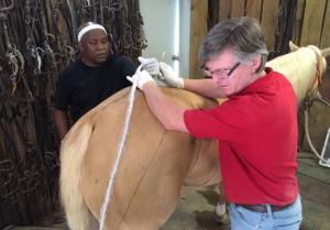
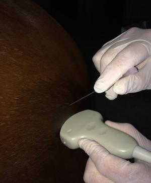
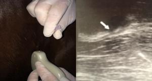
-Ultrasound guided SI injection with a combination of hyaluronic acid and steroids. In refractory cases, regenerative therapies such as IRAP, PRP, and stem cells have proven useful.
-Correcting any underlying primary issues which are causing the SI pain such as hock or stifle disease.
Trochanteric Bursitis
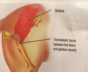
Anatomically, trochanteric bursitis (TB) is inflammation of the fluid of the middle gluteal muscle as it passes over the greater trochanter of the femur.
It is almost always secondary to hock and stifle disease much like SI issues. The lay term for this area is the Whorl Bone. When hock and stifle issues alter the horses correct “way of going”, inflammation in the bursa is common.
Clinical signs of TB (Whorl Bone lameness) include:
-pain on palpation of the area.
-a tendency to move away from the affected side on the straight line at a trot.
-Landing on the inside (medial) wall of the hoof first. This can sometimes be seen by excessive wear on the inside of the hind shoes only.
Treatment options are very similar to SI disease and include:
-rest.
-Correction of any predisposing hock or stifle issues.
-NSAIDS
-controlled exercise program
-ultrasound guided injection of the affected area with hyaluronic acid and steroid combination, IRAP, PRP or stem cells.
Cervical Arthropathy of the Neck
Within the cervical spine of the neck in the horse are small joints called facets. Although not commonly thought of, these are joints just like hocks, stifles, and ankles are joints. When these areas become painful or arthritic, they can create problems with performance and in some cases lameness.
Clinical signs of cervical issues include:
-pain or limited range of motion upon cervical flexion
-neurologic deficits causing mild ataxia or intermittent lameness in the front
-subtle lameness behind
-horses tend to be “luggy” and “lay in the bridle”.
Diagnosis is difficult and sometimes requires diagnostic aids such as radiography, ultrasound, nuclear scintigraphy (Bone scan) and myelograms.
Treatment of cervical vertebral arthritis includes:
-rest
-NSAIDS
-systemic steroids
-PSGAGS (Adequan)
-ultrasound guided cervical facet injection with Hyalurinic Acid/ Steroid combinations, PRP, IRAP, or Stem Cells.
As can be seen in these conditions, diagnosis can be difficult and treatment involves the utilization of different modalities all working together in conjunction with each other. The ultrasound guided technique should only be performed by an experienced Equine Practitioner with expertise in these procedures.
Dr. Akin is an Equine Practitioner in Collierville, TN whose practice is limited to lameness and performance issues in the Equine Athlete. Any questions concerning this article should be directed to Dr. Akin at MAkinDVM86@gmail.comor 901-854-6773 (901 85HORSE).
Today’s equine athlete is asked to perform at higher and higher levels. Despite great strides in Veterinary Medicine, nutrition, farriering, and conditioning, today’s equine athlete can become victim to subtle injury which can cause lameness, poor performance, or both. This article takes on three of those often overlooked causes of lameness and poor performance in an attempt to put them in perspective.
A Sacroiliac desmitis and arthropathy
B Trochanteric Bursitis
C Cervical Arthropathy
Sacroiliac Desmitis and Arthropathy
Anatomically the SI region pain is where the ilium (part of the pelvis) and the sacrum (part of the vertebral column) connect:
The Sacroiliac (SI) is often the culprit as it pertains to lower back issues, lameness and poor performance in both the English and Western performance horse. These SI can be primary or can be commonly secondary to lower limb issues concerning the hocks and stifles.
Clinical signs of sacroiliac disease in the performance horse include but aren’t limited to:
-poor performance
-unwillingness to go forward
-a refusal to “collect”
-pain on palpation of the lower back and croup (sacral tuberosity)
-kicking out when trotting or cantering
-poor attitude during exercise, just “crabby”
-frequent shifting of weight
-lack of desire to have rear flexions performed
Treatment involves multiple modalities and should be done in concert with each other. My typical treatment plan involves the following and can last from three weeks to 3 months.
-Rest. The cornerstone of any rehabilitation program.
-Controlled exercise program which involves a lot of lunging which leads to work under saddle. Starting with easy tasks for the horse and progressively asking for more over the rehab period.
-Non Steroidal Anti Inflammatory Drugs (NSAIDS), muscle relaxants, and ligament relaxers to break the chronic cycle of pain.
-Ultrasound guided SI injection with a combination of hyaluronic acid and steroids. In refractory cases, regenerative therapies such as IRAP, PRP, and stem cells have proven useful.
-Correcting any underlying primary issues which are causing the SI pain such as hock or stifle disease.
Trochanteric Bursitis
Anatomically, trochanteric bursitis (TB) is inflammation of the fluid of the middle gluteal muscle as it passes over the greater trochanter of the femur.
It is almost always secondary to hock and stifle disease much like SI issues. The lay term for this area is the Whorl Bone. When hock and stifle issues alter the horses correct “way of going”, inflammation in the bursa is common.
Clinical signs of TB (Whorl Bone lameness) include:
-pain on palpation of the area.
-a tendency to move away from the affected side on the straight line at a trot.
-Landing on the inside (medial) wall of the hoof first. This can sometimes be seen by excessive wear on the inside of the hind shoes only.
Treatment options are very similar to SI disease and include:
-rest.
-Correction of any predisposing hock or stifle issues.
-NSAIDS
-controlled exercise program
-ultrasound guided injection of the affected area with hyaluronic acid and steroid combination, IRAP, PRP or stem cells.
Cervical Arthropathy of the Neck
Within the cervical spine of the neck in the horse are small joints called facets. Although not commonly thought of, these are joints just like hocks, stifles, and ankles are joints. When these areas become painful or arthritic, they can create problems with performance and in some cases lameness.
Clinical signs of cervical issues include:
-pain or limited range of motion upon cervical flexion
-neurologic deficits causing mild ataxia or intermittent lameness in the front
-subtle lameness behind
-horses tend to be “luggy” and “lay in the bridle”.
Diagnosis is difficult and sometimes requires diagnostic aids such as radiography, ultrasound, nuclear scintigraphy (Bone scan) and myelograms.
Treatment of cervical vertebral arthritis includes:
-rest
-NSAIDS
-systemic steroids
-PSGAGS (Adequan)
-ultrasound guided cervical facet injection with Hyalurinic Acid/ Steroid combinations, PRP, IRAP, or Stem Cells.
As can be seen in these conditions, diagnosis can be difficult and treatment involves the utilization of different modalities all working together in conjunction with each other. The ultrasound guided technique should only be performed by an experienced Equine Practitioner with expertise in these procedures.
Dr. Akin is an Equine Practitioner in Collierville, TN whose practice is limited to lameness and performance issues in the Equine Athlete. Any questions concerning this article should be directed to Dr. Akin at MAkinDVM86@gmail.comor 901-854-6773 (901 85HORSE).


