
By Leigh Ballard
Hoof abscesses occur when bacteria become trapped under the hoof wall or sole. This can occur from several sources, but usually from a penetrating wound or foreign matter like soil or sand particles becoming trapped in a crack or defect in the white line and working its way into the sensitive structure of the hoof. Abscesses occur more often in the winter or spring after the horse has been subjected to wet and muddy conditions, causing his feet to be soft and vulnerable.
Trapped bacteria sets up infection, and the purulent fluid (pus) created builds up very painful pressure. The hoof wall can’t expand to ease the pressure, and it becomes so painful that the horse will not bear any weight on the foot. It is frightening to see a horse trying to walk with an abscess, because the onset is often sudden, and the horse who was fine yesterday is inexplicably “three-legged” lame today. The abscess can sometimes be located and drained by a veterinarian or farrier. Other times the problem is not so easy to find, and the abscess takes time to work itself out, often migrating to the coronary band where it finally exits the hoof.
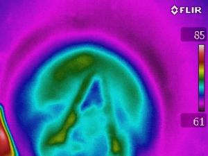
Below are some thermal images showing abscesses.
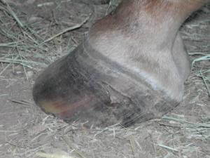
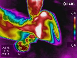
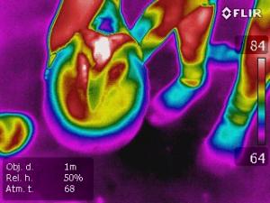
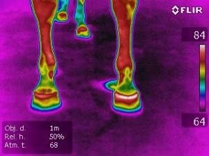
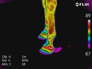
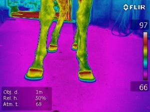
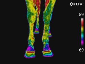
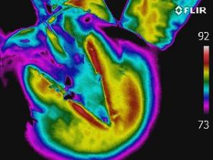
The first set of images [IR_0209, 0211, 0215, 0223] is of a broodmare heavy in foal, about 6 weeks from delivery. She was trimmed about 2 weeks prior to these images being taken. A few days after trimming she seemed a little off but, it seemed to resolved itself. Then another few days later she was off again, but not lame enough to think it was due to anything other than being heavily pregnant and a little tender footed. The veterinarian happened to visit the mare to give a vaccination during this episode, so hoof testers were used on her, but she had no response to those. Then, two days later she became unquestionably lame and these images were taken. The abscess finally exited at the coronary band after about a week of treatment. The last image shows the hoof about two months later.
Mare case study:
The second set of images [IR_0291, 0376, 0389, 0461] is of a gelding who came in from the pasture one day with a chip in his toe, but no lameness. After about two days he started showing signs of slight lameness that progressed into a more acute lameness a few days later. The images show a progression of the buildup of an abscess, from baseline taken prior, heat beginning to be evident in right toe, then when abscess was acute foot was cooled to determine exact spot of abscess. Baseline imaging of this horse had been done two weeks prior, so a normal front hoof view is included for comparison. This abscess was resolved by a farrier.
Images provided by Equine Imaging Solutions
Hoof abscesses occur when bacteria become trapped under the hoof wall or sole. This can occur from several sources, but usually from a penetrating wound or foreign matter like soil or sand particles becoming trapped in a crack or defect in the white line and working its way into the sensitive structure of the hoof. Abscesses occur more often in the winter or spring after the horse has been subjected to wet and muddy conditions, causing his feet to be soft and vulnerable.
Trapped bacteria sets up infection, and the purulent fluid (pus) created builds up very painful pressure. The hoof wall can’t expand to ease the pressure, and it becomes so painful that the horse will not bear any weight on the foot. It is frightening to see a horse trying to walk with an abscess, because the onset is often sudden, and the horse who was fine yesterday is inexplicably “three-legged” lame today. The abscess can sometimes be located and drained by a veterinarian or farrier. Other times the problem is not so easy to find, and the abscess takes time to work itself out, often migrating to the coronary band where it finally exits the hoof.
Below are some thermal images showing abscesses.
The first set of images [IR_0209, 0211, 0215, 0223] is of a broodmare heavy in foal, about 6 weeks from delivery. She was trimmed about 2 weeks prior to these images being taken. A few days after trimming she seemed a little off but, it seemed to resolved itself. Then another few days later she was off again, but not lame enough to think it was due to anything other than being heavily pregnant and a little tender footed. The veterinarian happened to visit the mare to give a vaccination during this episode, so hoof testers were used on her, but she had no response to those. Then, two days later she became unquestionably lame and these images were taken. The abscess finally exited at the coronary band after about a week of treatment. The last image shows the hoof about two months later.
Mare case study:
The second set of images [IR_0291, 0376, 0389, 0461] is of a gelding who came in from the pasture one day with a chip in his toe, but no lameness. After about two days he started showing signs of slight lameness that progressed into a more acute lameness a few days later. The images show a progression of the buildup of an abscess, from baseline taken prior, heat beginning to be evident in right toe, then when abscess was acute foot was cooled to determine exact spot of abscess. Baseline imaging of this horse had been done two weeks prior, so a normal front hoof view is included for comparison. This abscess was resolved by a farrier.
Images provided by Equine Imaging Solutions


