By R. M. Bowker,VMD, PHD
Department of Pathobiology and Diagnostic Investigation, Michigan State University
The horse’s foot is the primary avenue for the horse to obtain information about the physical features of the ground and environment. The horse gains access to this information via the multitude of sensory nerves and receptors that are distributed throughout the foot. Activation of these sensory receptors allows the information to be incorporated into different reflexes needed for movement, posture, and/or protection. While veterinary medicine has focused more on the pain-carrying nerve fibers, horses have other sensations that are so important in their daily lives. These sensations are detected by mechanoreceptors—sensory structures that are activated by physical deformation rather than destruction of tissue needed for pain. Most of the sensations from the mechanoreceptors are involved in reflexes, with some also going to brain for conscious perception by the horse. The reflex inputs go to the Spinal Cord Generator (SCG), which enable the coordination of the movements of all limbs and back muscles during athletic performances and stance. The activation of these sensory mechanoreceptors have widespread effects over the entire body: they aid in permitting intricate coordination of the long muscles along the back and neck with the feet, and even the head and teeth as well as produce local effects within the tissues by improving blood perfusion through the foot.
So what are these sensory receptors within the horse’s foot that are crucial for the horse’s perception of its environment? The feet of most mammals have contact with the ground via their hooves and/or foot pads, which protect the inner workings of the foot or fingers while the internal tissues detect and perceive the environmental stimuli. These tissues protect the foot as well as provide a means to obtain information about its surrounding environment. The mechanoreceptors convey sensations of touch, pressure, and vibration in the daily activities of the horse. Which specific mechanoreceptors are activated depend on which foot structures are loaded at any given time. Changes in hoof balance, shoeing, and ground surface features all affect the sensations that the horse perceives.
There are three categories of these mechanoreceptors:light cutaneous contact or touch receptors, which are located superficially in the skin of the coronet or frog, and the hoof wall; deep pressure receptors, whichare excited when the foot tissues are fully loaded during stance or movement; the third group perceive vibrations. Two of these sensory receptors are particularly interesting physiologically as they enable the horse to (1) feel its environment and (2) to detect vibrations during movements or tremors from the ground: (1) Merkel's discs (and touch receptors) and(2)Pacinian corpuscles.
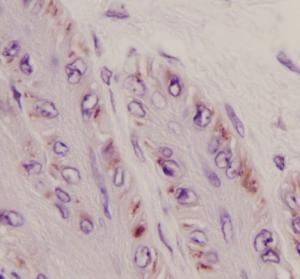
Within the skin overlying the heel bulbs and coronet, many free nerve endings are present. They detect touch/light pressure sensations applied to the skin, usually caused by insects, grasses, etc. and during foot loading, thermal sensations as well. When a hair is bent, the underlying hair follicle stretches and send the impulses to the spinal cord. The hair shaft enhances the sensitivity when it is touched. Merkel’s discs (figure) are present within the deeper layers of the skin and hoof wall and respond to continuously applied light touch and pressure. These receptors on the sole and frog may be activated by a terry cloth, as the horse feels more comfortable standing on the cloth than on a hard surface!
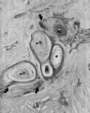
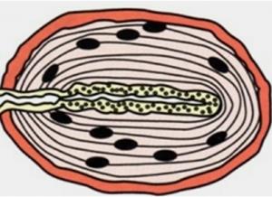
During foot loading, the Pacinian corpuscles become activated to provide critical information for movement. These very large receptors are shaped like an onion and in several locations in the foot, including the bulbs of heel (Figure), surrounding the frog near the central sulcus, in the digital cushion, and near the insertion of the deep digital flexor tendon (DDFT) onto short pastern bone. These are critical for providing sensory information from the foot during ground contact. This sensory information goes to the SCG, enabling flexor and extensor muscles to function in a coordinated fashion as the horse moves across varying ground surfaces. Gait abnormalities may result in changes in the sensory inputs to limb muscles from these receptors. A toe-stabbing gait could be a mismatch between Pacinian corpuscle and touch input to the SCG. Sensory perception would likely be altered and produce an abnormal foot fall, leading to stumbling. Horses with unbalanced feet (“high-low” syndrome or a “club” foot) may have altered sequencing of the receptors and limb muscles, resulting in changing muscle contour at the shoulder, as do long-toed and under-run heeled horses.
Metal-shod horses have greater impact energies passing through the foot to these mechanoreceptors, and the increased high energy frequency waves mayaffect the sensitivity of these receptors during movements. Pacinian corpuscles (Figure drawing) are sensitive to low frequency vibratory signals (infrasounds; 20-50 Hz) during stance. Their locations in the frog and heel bulbs indicate that they are poised to receive vibrations and tremors through the ground. Tremors are often detected days in advance of a surface eruption of an earthquake. Perhaps the uneasiness of horses and other farm animals several days in advance of an earthquake may be due to sensing these underground tremors. Other observations suggest that these vibration-responsive corpuscles may be detecting the reflected sound waves from large rocks and boulders deep in the ground that are produced by trotting or galloping horses. Anecdotal observations mention that galloping horses begin to slow down as they approach areas where underground terrain changes, suggesting that these waveforms may be detectable when the horses are moving over the ground surfaces. In any event, horses appear to be able to “hear” with their feet!
The close relationship of the Pacinian corpuscles and the openings of the scent glands onto the frog central sulcus suggests that foot loading also has a “neuroexocrine” function. While sensory information is transmitted to the spinal cord for reflex coordination of limbs during this time of foot contact with the ground, the scent glands opening onto the central sulcus deposit secretions on the ground, enabling communication between horses through the sense of smell. This may explain why horses in a herd often smell the ground where the dominant horse has walked.
The clinical significance of these receptors of the skin and foot is that they provide the horse with critical information about its environment for both movement and stance. During gait abnormalities, some of this sensory information is “deleted” from the SCG, resulting in impaired motor pattern messages to the limb muscles. When receptors and spinal reflexes provide improper information to the SCG, then the result is abnormal limb and foot mechanics. To the observer it appears that the horse has forgotten how to move or stand normally, or may move clumsily. While traditional diagnostics often reveal a horse to be within normal limits, despite performance complaints by the owners, other methods, including acupuncture, chiropractic, or physical therapy may be needed to re-engage these sensory receptors of the feet and body to assist return to normal sensory and motor function. Rehabilitation serves to normalize the SCG neuronal circuits to the appropriate movement pattern. When abnormal movement patterns have become habitual, the nervous system adapts and undergoes re-programming of the SCG to accommodate the abnormal postural stance and movement patterns. In this case the mechanoreceptors not only in the feet, but all over the body must be re-engaged to return the equine athletes to their high levels of performance. When such coordinated neuromotor activity and sensory integration occurs, the result is what owners wish to see in their horses: coordinated, graceful, efficient movement as the horse engages in work under saddle or freely moving in pastures. From the horse’s perspective, the horse feels its environment better and may seek out areas in the barnyard and pasture that are more comfortable for him.
Photos: Pacinian-1: picture of pacinian corpuscle in heel bulbs of foot; shows several onion shaped structures.


